


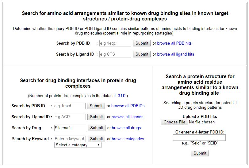
To provide examples on how the web server can be used for protein binding site prediction, we illustrate some case studies used to analyse protein-drug interactions in different perspectives.
Sildenafil is a phosphodiesterase (PDE) inhibitor, and is known to bind to phosphodiesterase 5 (PDE5). The drug has previously been prescribed for pulmonary arterial hypertension (PAH), but later it had been repurposed for erectile dysfunction.
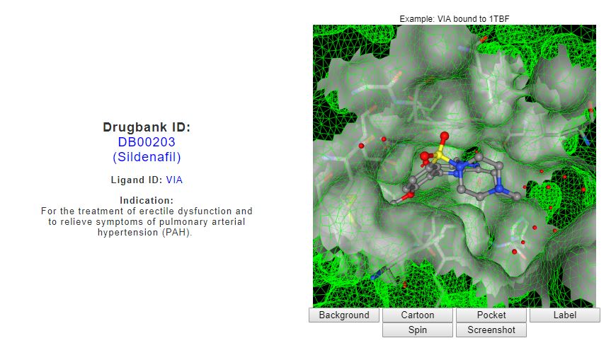
From the ‘Search for drug binding interfaces in protein-drug complexes’ interface, drug search for ‘Sildenafil’ returns the drug indication as retrieved from the Drugbank, a visualization of PDB structure for Sildenafil molecule (PDB ligand ID: VIA), and a list of known binding sites for Sildenafil derived from the PDB.
(Link)
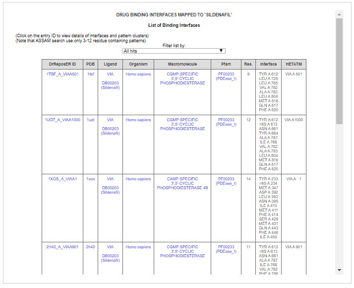
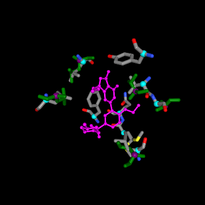
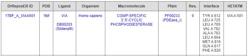
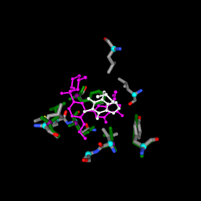
Sildenafil are found in complex with different PDB structures of human cGMP-specific 3’,5’-cyclic phosphodiesterase. One of them is a phosphodiesterase 5A (PDB ID: 1tbf) that contains 9-residue pattern surrounds a Sildenafil molecule.
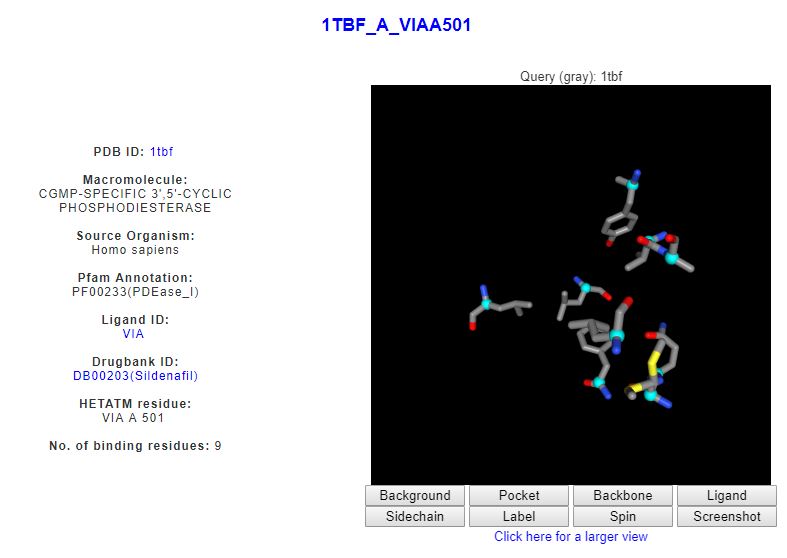
Clicking on the first DrReposer ID '1TBF_A_VIAA501' returns a new page for similar patterns of amino acids predicted from the ASSAM search. User may sort the results according to RMSD, Z-score or sequence identity values.
(Link)
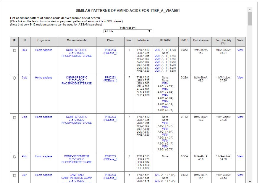
Some of the hits include related phosphodiesterases. One of the hits is a cAMP-specific 3’,5’-cyclic phosphodiesterase 7A (PBD ID: 1zkl) with high fold similarity to the query protein (PDB ID: 1tbf).

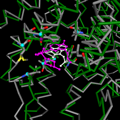
ASSAM search found matched 5 residues that are similarly arranged as the query binding site and the residues are bound to 3-isobutyl-1-methylxanthine, an experimental small molecule (PDB ligand ID: IBM).
Upon clicking the ‘View’ link on the last column, matched pattern of amino acids is shown in green, which is superposed to the query binding site (grey). Superposed ligand can also be seen, i.e. hit ligand is indicated in white, by clicking the 'Ligand' button on the NGL viewer. Ligand ID IBM can be seen to be superposed with Sildenafil molecule.
Superposition of backbone structure can also be seen which allow user to inspect the structural similarity.
Another interesting hit a matched 5-residue pattern in an uncharacterized protein from Mycobacterium tuberculosis (PBD ID: 4nxi) with no similarity in terms of sequence or fold to the query protein.